Bassett Collection of Stereoscopic Images of Human Anatomy
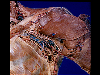
Pectoral region and axilla
Left axilla; coracoclavicular fascia removed
Image #90-2
KEYWORDS: Axilla, Peripheral nervous system.
Creative Commons
Stanford holds the copyright to the David L. Bassett anatomical images and has assigned Creative Commons license Attribution-Share Alike 4.0 International to all of the images.

For additional information regarding use and permissions, please contact the Medical History Center.


Pectoral region and axilla
Left axilla; coracoclavicular fascia removed
The coracoclavicular fascia (12) has been partially removed and the connective tissue taken out of the axillary fossa.
- Trapezius muscle
- Articular branch of lateral anterior thoracic nerve to acromioclavicular and shoulder joints
- Clavicle
- Subclavius muscle
- Anterior lateral thoracic nerve
- Lateral cord of brachial plexus
- Axillary artery and vein
- Subclavian trunk
- Axillary lymph nodes (subclavian group)
- Axillary lymph nodes (central group)
- Rib II
- Coracoclavicular fascia
- Intercostobrachial nerve
- Axillary lymph nodes (anterior pectoral group)
- Deltoid muscle
- Thoracoacromial artery (cut off)
- Pectoralis minor muscle (reflected laterally)
- Anterior medial thoracic nerve
- Supreme thoracic artery
- Pectoralis major muscle (reflected laterally)
- Axillary lymph nodes (lateral group)
- Upper pointer: Axillary fascia Lower pointer: Lateral thoracic artery