Bassett Collection of Stereoscopic Images of Human Anatomy
Pectoral region and axilla
Right axillary fascia, inferior aspect
Image #90-1
KEYWORDS: Axilla, Fascia ligaments and tendons, Pectoral region.
Creative Commons
Stanford holds the copyright to the David L. Bassett anatomical images and has assigned Creative Commons license Attribution-Share Alike 4.0 International to all of the images.
For additional information regarding use and permissions, please contact the Medical History Center.
Pectoral region and axilla
Right axillary fascia, inferior aspect
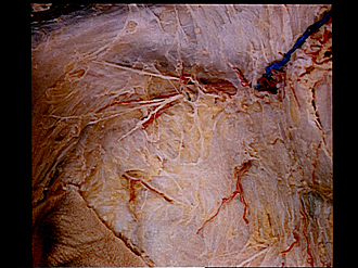
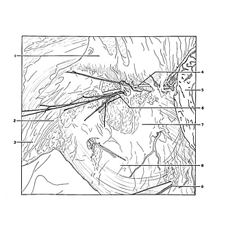
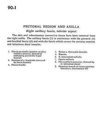
The skin and subcutaneous connective tissue have been removed from the right axilla. The axillary fascia (7) is continuous with the pectoral (1) and brachial fascia (3) and with the fascia which covers the serratus anterior and latissimus dorsi muscles.
- Pectoral fascia (pointer on anterior axillary fold, formed by underlying pectoralis major muscle)
- Position of brachial artery (covered by brachial fascia)
- Brachial fascia
- Branch lateral thoracic artery
- Breast
- Intercostobrachial nerve
- Axillary fascia
- Posterior axillary fold (formed by latissimus dorsi muscle)
- Posterior branch of lateral cutaneous branch of intercostal nerve III


