Bassett Collection of Stereoscopic Images of Human Anatomy
Pectoral region and axilla
Left thoracoacromial vessels
Image #89-5
KEYWORDS: Pectoral region, Peripheral nervous system, Vasculature.
Creative Commons
Stanford holds the copyright to the David L. Bassett anatomical images and has assigned Creative Commons license Attribution-Share Alike 4.0 International to all of the images.

For additional information regarding use and permissions, please contact the Medical History Center.

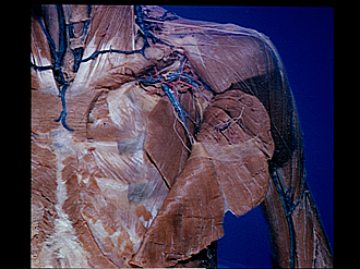
Pectoral region and axilla
Left thoracoacromial vessels
The pectoralis major muscle has been reflected inferiorly from its origin. The fascia which covered the deep surface of the muscle has been removed except for a narrow band (18). The fascia has also been removed from the pectoralis minor muscle (7) and this muscle divided but not reflected.
- External jugular vein
- Middle supraclavicular nerves
- Sternocleidomastoid muscle
- Upper pointer: Clavicular branch of thoracoacromial artery Lower pointer: Axillary lymph node (of subclavian group)
- Thoracoacromial vein
- Branches of anterior lateral thoracic nerve
- Pectoralis minor muscle
- Rib II
- Body of sternum
- Sternocostal origins of pectoralis major muscle (cut off)
- Deltoid muscle
- Upper pointer: Clavicle Lower pointer: Subclavius muscle
- Acromial branch thoracoacromial artery
- Anterior lateral thoracic nerve (cut off)
- Branch to deltoid of thoracoacromial artery
- Anterior lateral thoracic nerve (distal continuation of 14)
- Pectoral branch of thoracoacromial artery
- Pectoral fascia
- Pectoralis major muscle
- Cephalic vein