Bassett Collection of Stereoscopic Images of Human Anatomy
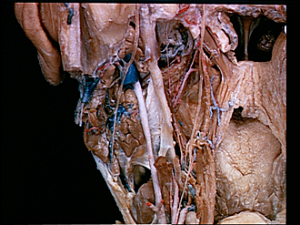
Dissection of head and neck from a posterior approach
Relation of left facial nerve to parotid gland
Image #83-1
KEYWORDS: Bones cartilage joints, Cheek, Exocrine and endocrine, Face, Mouth, Peripheral nervous system.
Creative Commons
Stanford holds the copyright to the David L. Bassett anatomical images and has assigned Creative Commons license Attribution-Share Alike 4.0 International to all of the images.

For additional information regarding use and permissions, please contact the Medical History Center.


Dissection of head and neck from a posterior approach
Relation of left facial nerve to parotid gland
The sternocleidomastoid muscle (3) has been cut off close to its insertion and its tendon reflected laterally from the mastoid process (4) of the temporal bone. The posterior facial vein (7) has been cut off and the parotid gland dissected to expose the facial nerve as it passes between the deep (9) and superficial (10) lobes of the gland.
- Stylomastoid foramen (opened)
- Mastoid incisure
- Sternocleidomastoid muscle (tendon of insertion stripped from mastoid process)
- Mastoid process
- Facial nerve (VII)
- Superior (temporofacial) division of facial nerve (VII)
- Posterior facial vein
- Inferior (cervicofacial) division of facial nerve (VII)
- Parotid gland (deep lobe)
- Parotid gland (superficial lobe)
- External carotid artery
- Stylohyoid ligament
- Stylohyoid muscle (cut across)
- Upper pointer: Hypoglossal nerve (XII) (cut across) Lower pointer: External maxillary artery
- Superficial fascia (external layer)
- Submandibular gland
- Mandibular nerve
- External pterygoid muscle
- Styloid process temporal bone
- Salpingopharyngeus muscle
- Upper pointer: Ascending palatine artery Lower pointer: Stylomandibular ligament
- Superior pharyngeal constrictor muscle (pointer near origins of muscle from pterygomandibular raphe)
- Styloglossus muscle
- Glossopharyngeal nerve (IX)
- Stylopharyngeus muscle (cut across)