Bassett Collection of Stereoscopic Images of Human Anatomy
Dissection of anterior and lateral regions of neck
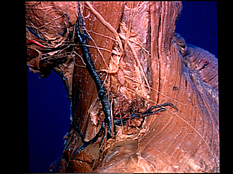
Contents of left posterior cervical triangle; superficial cervical lymphatics, lateral view
Image #73-1
KEYWORDS: Fascia and connective tissue, Lymphatics, Muscles and tendons.
Creative Commons
Stanford holds the copyright to the David L. Bassett anatomical images and has assigned Creative Commons license Attribution-Share Alike 4.0 International to all of the images.

For additional information regarding use and permissions, please contact the Medical History Center.


Dissection of anterior and lateral regions of neck
Contents of left posterior cervical triangle; superficial cervical lymphatics, lateral view
The external layer of cervical fascia has been removed from the left side of the neck except from the lower part of the posterior triangle. In this region the lamina of this fascia which passes deep to the sternocleidomastoid and trapezius muscles remains in place. This layer has openings for lymphatic vessels (20) and veins, but can be traced superiorly to the midpart of the view where it becomes continuous with the prevertibral fascia (13). The left shoulder occupies the midforeground of the view.
- Upper pointer: Parotid gland Lower pointer: Parotid-masseteric fascia
- Cervical branch of facial nerve
- Mandibular marginal branch of facial nerve
- Anastomosis of cervical cutaneus nerve with facial nerve
- Platysma (reflected anteriorly)
- External jugular vein
- Cutaneus colli nerve
- Sternocleidomastoid muscle
- Anterior supraclavicular nerves
- Medial supraclavicular nerves
- Upper pointer: Clavicle (acromial end) Lower pointer: Deltoid muscle
- Great auricular nerve
- Prevertebral fascia
- Minor occipital nerve
- Borders of posterior cervical triangle
- Accessory nerve (XI)
- Trapezius muscle
- Posterior supraclavicular nerves
- Superficial cervical lymph glands
- Superficial lymphatic vessel passing through cervical fascia
- [Legend above restored translation from Latin]