Bassett Collection of Stereoscopic Images of Human Anatomy
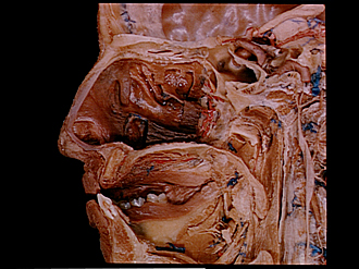
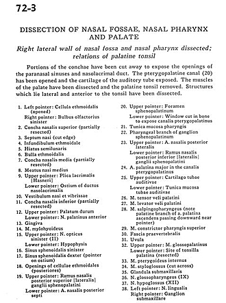
Dissection of nasal fossae, nasal pharynx, and palate
Right lateral wall of nasal fossa and nasal pharynx dissected; relations of palatine tonsil
Image #72-3
KEYWORDS: Face, Mouth, Nose.
Creative Commons
Stanford holds the copyright to the David L. Bassett anatomical images and has assigned Creative Commons license Attribution-Share Alike 4.0 International to all of the images.

For additional information regarding use and permissions, please contact the Medical History Center.
Dissection of nasal fossae, nasal pharynx, and palate
Right lateral wall of nasal fossa and nasal pharynx dissected; relations of palatine tonsil
Portions of the conchae have been cut away to expose the openings of the paranasal sinuses and nasolacrimal duct. The pterygopalatine canal (20) has been opened and the cartilage of the auditory tube exposed. The muscles of the palate have been dissected and the palatine tonsil removed. Structures which lie lateral and anterior to the tonsil have been dissected.
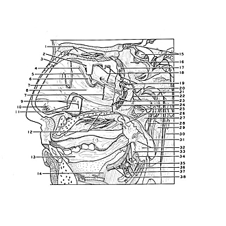
- Left pointer: Ethmoidal cell (opened) Right pointer: Olfactory bulb left
- Superior nasal concha (partially resected)
- Nasal septum (cut edge)
- Ethmoidal infundibulum
- Hiatus semilunaris
- Ethmoidal bulla
- Middle nasal concha (partially resected)
- Middle nasal meatus
- Upper pointer: Lacrimal fold Lower pointer: Ostium of nasolacrimal duct
- Vestibule nasi and vibrissae (hair)
- Inferior nasal concha (partially resected)
- Upper pointer: Hard palate Lower pointer: Anterior palatine nerve
- Gingiva
- Mylohyoid muscle
- Upper pointer: Optic nerve (II) left Lower pointer: Hypophysis
- Sphenoid sinus left
- Sphenoid sinus right (pointer on ostium)
- Openings of ethmoidal cells (posterior)
- Upper pointer: Posterior superior nasal branch of sphenopalatine ganglion Lower pointer: Posterior nasal septal artery
- Upper pointer: Sphenopalatine foramen Lower pointer: Window cut in bone to expose pterygopalatine canal
- Tunica mucosa pharynx
- Pharyngeal branch of sphenopalatine ganglion
- Upper pointer: Posterior lateral nasal artery Lower pointer: Posterior inferior (lateral) nasal branch of sphenopalatine ganglion
- Greater palatine artery in the pterygopalatine canal
- Upper pointer: Cartilaginous auditory tube Lower pointer: Mucosa
- Palatine tensor veli muscle
- Palatine levator veli muscle
- Salpingopharyngeal muscle (note palatine branch of ascending palatine artery passing downward near pointer)
- Superior constrictor pharyngis muscle
- Prevertebral fascia
- Uvula
- Upper pointer: Glossopalatine muscle Lower pointer: Site of palatine tonsilla (resected)
- Internal pterygoid muscle
- Styloglossus muscle (cut across)
- Submaxillary gland
- Glossopharyngeal muscle (IX)
- Hypoglossus nerve (XII)
- Left pointer: Lingual nerve Right pointer: Submaxillary ganglion
- [Legend above restored translation from Latin]