Bassett Collection of Stereoscopic Images of Human Anatomy
Oral cavity
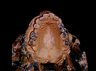
Roof of oral cavity, inferior view
Image #70-6
KEYWORDS: Bones cartilage joints, Face, Mouth.
Creative Commons
Stanford holds the copyright to the David L. Bassett anatomical images and has assigned Creative Commons license Attribution-Share Alike 4.0 International to all of the images.
For additional information regarding use and permissions, please contact the Medical History Center.
Oral cavity
Roof of oral cavity, inferior view
The palatine arches (13,14) have been cut across and the tongue, mandible and associated structures have been removed. The left upper lip and buccal wall have been cut away. The teeth are numbered in the drawing in the conventional fashion. The third molar teeth were not present.
- Medial incisor
- Lateral incisor
- Canine
- Premolar I
- Premolar II
- Molar I
- Molar II
- Upper lip
- Angle of mouth
- Buccinator muscle (at labial commissure)
- Hard palate
- Gingiva
- Upper pointer: Glossopalatine muscle (cut off) Lower pointer: Palatoglossal arch
- Upper pointer: Palatopharyngeal arch Lower pointer: Palatopharyngeal muscle
- Incisive papilla
- Transverse palatine fold
- Palatine raphe
- Palatine fovea
- Soft palate
- Uvula
- Palatine tonsil (atrophic)


