Bassett Collection of Stereoscopic Images of Human Anatomy
Dissection of left parotideomasseteric region
Facial nerve within parotid gland
Image #63-3
KEYWORDS: Cheek, Exocrine and endocrine, Face, Peripheral nervous system.
Creative Commons
Stanford holds the copyright to the David L. Bassett anatomical images and has assigned Creative Commons license Attribution-Share Alike 4.0 International to all of the images.
For additional information regarding use and permissions, please contact the Medical History Center.
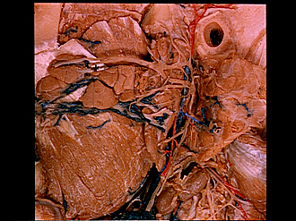
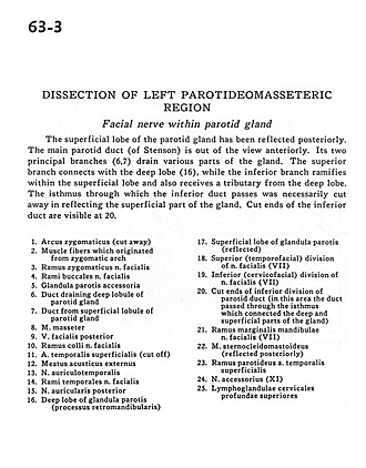
Dissection of left parotideomasseteric region
Facial nerve within parotid gland
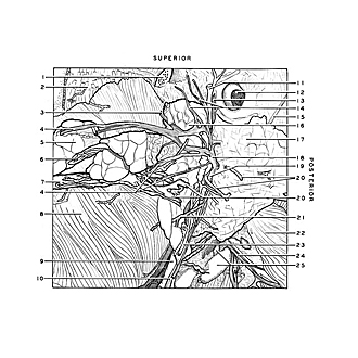
The superficial lobe of the parotid gland has been reflected posteriorly. The main parotid duct (of Stenson) is out of view anteriorly. Its two principal branches (6,7) drain various parts of the gland. The superior branch connects with the deep lobe (16), while the inferior branch ramifies within the superficial lobe and also receives a tributary from the deep lobe. The isthmus through which the inferior duct passes was necessarily cut away in reflecting the superficial part of the gland. Cut ends of the inferior duct are visible at 20.
- Zygomatic arch (cut away)
- Muscle fibers which originated from zygomatic arch
- Zygomatic branches of facial nerve
- Buccal branches of facial nerve
- Accessory parotid gland
- Duct draining deep lobule of parotid gland
- Duct from superficial lobe of parotid gland
- Masseter muscle
- Posterior facial vein
- Superficial branch facial nerve
- Superficial temporal artery (cut off)
- External acoustic meatus
- Auriculotemporal nerve
- Temporal branches of facial nerve
- Posterior auricular nerve
- Deep lobe of parotid gland (retromandibular process)
- Superficial lobe of parotid gland (reflected)
- Superior (temporofacial) division of facial nerve (VII)
- Inferior (cervicofacial) division of facial nerve (VII)
- Cut ends of inferior division of parotid duct (in this area the duct passed through the isthmus which connected the deep and superficial parts of the gland)
- Marginal mandibular branch facial nerve (VII)
- Sternocleidomastoid muscle (reflected posteriorly)
- Parotid branch superficial temporal artery
- Accessory nerve (XI)
- Superior deep cervical lymph nodes


