Bassett Collection of Stereoscopic Images of Human Anatomy
Dissection of right orbit from a superior approach
General relations of structures within orbit; orbital fascia
Image #54-1
KEYWORDS: Connective tissue, Eye, Face, Muscles and tendons.
Creative Commons
Stanford holds the copyright to the David L. Bassett anatomical images and has assigned Creative Commons license Attribution-Share Alike 4.0 International to all of the images.

For additional information regarding use and permissions, please contact the Medical History Center.

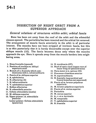
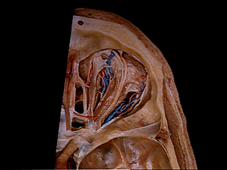
Dissection of right orbit from a superior approach
General relations of structures within orbit; orbital fascia
Bone has been cut away from the roof of the orbit and the ethmoidal sinuses opened. The periorbita has been resected and the orbital fat removed. The arrangement of muscle fascia anteriorly in the orbit is of particular interest. The muscles have not yet been stripped of intrinsic fascia, but this is so thin posteriorly that it is barely discernable except over the superior oblique muscle (12). The fascia becomes dense only where the muscles approach the eye. Here it spreads away from the muscle borders into neighboring areas.
- Frontal sinus (opened)
- Position of superior oblique muscle
- Branch of frontal nerve which communicates with infratrochlear nerve
- Fascia of superior oblique muscle
- Ophthalmic artery
- Olfactory nerve (I)
- Infratrochlear nerve
- Superior ophthalmic vein
- Olfactory bulb
- Anterior ethmoidal nerve
- Ethmoidal air cell (medial)
- Superior oblique muscle
- Upper pointer: Posterior ethmoidal artery Lower pointer: Ethmoidal cell (posterior)
- Trochlear nerve (IV)
- Roof of optic canal (upper root of lesser wing of sphenoid bone
- Dura mater and optic nerve (II)
- Anterior clinoid process
- Supratrochlear nerve
- Frontal nerve (supraorbital and frontal branches not separated)
- Fascia of levator palpebrae superioris muscle
- Levator palpebrae superioris muscle
- Fascia of superior rectus muscle
- Periorbita
- Superior rectus muscle
- Lacrimal nerve, artery, and vein
- Temporalis muscle
- Superior ophthalmic vein
- Frontal nerve