Bassett Collection of Stereoscopic Images of Human Anatomy
Exploration of the brain from its basal aspect
Ansa lenticularis
Image #5-7
KEYWORDS: Brain, Midbrain, Telencephalon.
Creative Commons
Stanford holds the copyright to the David L. Bassett anatomical images and has assigned Creative Commons license Attribution-Share Alike 4.0 International to all of the images.

For additional information regarding use and permissions, please contact the Medical History Center.


Exploration of the brain from its basal aspect
Ansa lenticularis
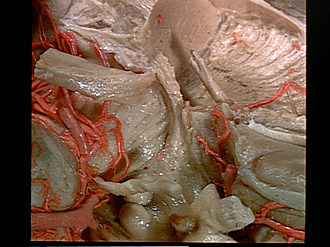
This view is a close-up of the previous one but the specimen is now turned at right angles so that the midline is at the bottom. Details of the ansa lenticularis (13) in its course around the medial edge of the cerebral peduncle are revealed. Some fibers of this group are also seen as they pass inward between fascicles of the peduncle and these join others above the peduncle to form the fasciculus lenticularis (H2 field of Forel) in the subthalamic region.
- Lateral geniculate body
- Optic tract (cut and retracted posteriorly)
- Cerebral peduncle
- Posterior cerebral artery (cut across)
- Pons
- Superior cerebellar artery
- Oculomotor nerve (III)
- Mamillary body
- Putamen
- Internal capsule
- Globus pallidus (external division)
- Anterior commissure
- Ansa lenticularis
- Artery of Heubner
- Diagonal band of Broca
- Radiating fibers of rostral lamina of corpus callosum
- Anterior perforated substance
- Anterior cerebral artery (cut across)
- Olfactory trigone
- Optic nerve (II)