Bassett Collection of Stereoscopic Images of Human Anatomy
Osteology
Roentgenogram of skull, inferosuperior view
Image #35-7
KEYWORDS: Bones cartilage joints, Overview.
Creative Commons
Stanford holds the copyright to the David L. Bassett anatomical images and has assigned Creative Commons license Attribution-Share Alike 4.0 International to all of the images.

For additional information regarding use and permissions, please contact the Medical History Center.
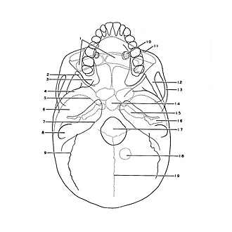
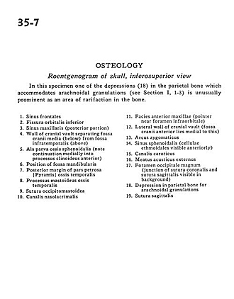
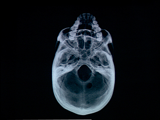
Osteology
Roentgenogram of skull, inferosuperior view
In this specimen one of the depressions (18) in the parietal bone which accommodates arachnoidal granulations (see Section I, 1-3) is unusually prominent as an area of rarification in the bone.
- Frontal sinus
- Inferior orbital fissure
- Maxillary sinus (posterior portion)
- Wall of cranial vault separating middle cranial fossa (below) from infratemporal fossa (above)
- Lesser wing of sphenoid bone (note continuation medially into anterior clinoid process)
- Position of mandibular fossa
- Posterior margin of petrosal part of temporal bone
- Mastoid process
- Occipitomastoid suture
- Nasolacrimal canal
- Anterior surface of maxilla (pointer near Infraorbital foramen)
- Lateral wall of cranial vault (anterior cranial fossa lies medial to this)
- Zygomatic arch
- Sphenoid sinus (ethmoidal cells visible anteriorly)
- Carotid canal
- External acoustic meatus
- Foramen magnum occipital (junction of coronal suture and sagittal suture visible in background)
- Depression in parietal bone for arachnoidal granulations
- Sagittal suture