Bassett Collection of Stereoscopic Images of Human Anatomy
Serial transverse sections of the brain stem
Medulla oblongata.
Image #30-1
KEYWORDS: Brain, Medulla, Midbrain, Peripheral nervous system, Pons.
Creative Commons
Stanford holds the copyright to the David L. Bassett anatomical images and has assigned Creative Commons license Attribution-Share Alike 4.0 International to all of the images.
For additional information regarding use and permissions, please contact the Medical History Center.
Serial transverse sections of the brain stem
Medulla oblongata.
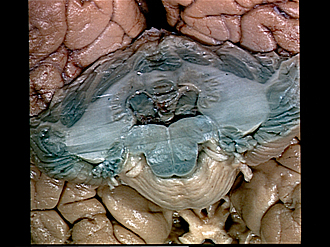
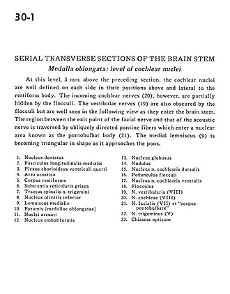
At this level, 3 mm. above the preceding section, the cochlear nuclei are well defined on each side of their positions above and lateral to the restiform body. The incoming cochlear nerves (20), however, are partially hidden by the flocculi. The vestibular nerves (19) are also obscured by the flocculi but are well seen in the following view as they enter the brain stem. The region between the exit point of the facial nerve and that of the acoustic nerve is transversed by obliquely directed pontine fibers which enter a nuclear area known as the pontobulbar body (21). The medial lemniscus (9) is becoming triangular in shape as it approaches the pons.
- Dentate nucleus
- Medial longitudinal fasciculus
- Choroid plexus fourth ventricle
- Area acustica
- Restiform body (inferior cerebellar peduncle)
- Reticular gray matter
- Spinal trigeminal tract
- Inferior olivary nucleus
- Medial lemniscus
- Pyramid (medulla oblongata)
- Arcuate nuclei
- Emboliform nucleus
- Globose nucleus
- Nodulus
- Dorsal cochlear nucleus
- Floccular peduncle
- Ventral cochlear nucleus
- Flocculus
- Vestibulocochlear nerve (VIII) vestibular part
- Vestibulocochlear nerve (VIII) cochlear part
- Facial nerve (VII) and "corpus pontobulbare"
- Trigeminal nerve (V)
- Optic chiasm


