Bassett Collection of Stereoscopic Images of Human Anatomy
Serial transverse sections of the brain stem
Medulla oblongata.
Image #29-6
KEYWORDS: Brain, Medulla, Midbrain.
Creative Commons
Stanford holds the copyright to the David L. Bassett anatomical images and has assigned Creative Commons license Attribution-Share Alike 4.0 International to all of the images.
For additional information regarding use and permissions, please contact the Medical History Center.
Serial transverse sections of the brain stem
Medulla oblongata.
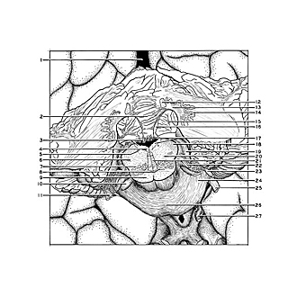
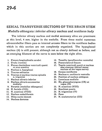
The inferior olivary nucleus and medial accessory olive are prominent at this level, 4 mm. higher in the medulla. From these nuclei numerous olivocerebellar fibers pass as internal arcuate fibers to the restiform bodies which in this section are not completely organized. The hypoglossal nucleus (4) is still present, although not as clearly defined as before, and an emerging filament of the nerve is seen below the right olive.
- Longitudinal fissure (cerebral)
- Uvula (vermis)
- Choroid plexus fourth ventricle and area acustica
- Nucleus hypoglossal nerve (XII).
- Tractus solitarius
- Spinal trigeminal tract and nucleus
- Inferior olivary nucleus
- Medial accessory olivary nucleus
- Pyramid (medulla oblongata)
- Facial nerve (VII)
- Vestibulocochlear nerve (VII)
- Emboliform nucleus
- Hilus dentate nucleus
- Dentate nucleus
- Tonsil (ventral paraflocculus)
- Posterolateral fissure
- Taenia fourth ventricle and dorsal motor nucleus of vagus nerve (X) (dorsal motor nucleus of the vagus nerve)
- Restiform body (inferior cerebellar peduncle )
- Ventral cochlear nucleus
- Position of nucleus ambiguous
- Medial lemniscus
- Glossopharyngeal nerve (IX) and vagus (X)
- Vestibulocochlear nerve (VIII)
- Middle cerebellar peduncle
- Trigeminal nerve (V)
- Pons
- Oculomotor nerve (III)


