Bassett Collection of Stereoscopic Images of Human Anatomy
Serial transverse sections of the brain stem
General orientation view.
Image #29-1
KEYWORDS: Brain, Cerebellum, Medulla, Midbrain, Pons, Telencephalon, Overview.
Creative Commons
Stanford holds the copyright to the David L. Bassett anatomical images and has assigned Creative Commons license Attribution-Share Alike 4.0 International to all of the images.
For additional information regarding use and permissions, please contact the Medical History Center.
Serial transverse sections of the brain stem
General orientation view.
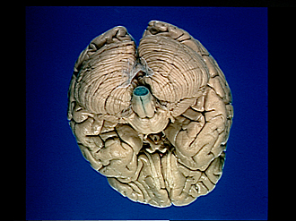
In this series of views of the brain, the brain stem has been cut transversely at successively higher levels and the cut surfaces stained with a dilute alcoholic solution of nile blue sulphate. By this means the myelin sheaths of nerve fibers become stained blue in contrast to gray matter which remains pale. (Note: connective tissue and blood vessels take on a dark color also.) In a general way the staining characteristics of the cut surface resemble those seen in sections stained by the Weigert method for microscopic study. Although detail here is diminished by the opacity of the tissue, the major features of the cut surface at each level are readily distinguished and these characteristics can be related to the gross features of the brain.
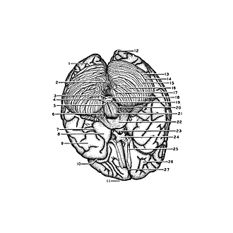
- Transverse cerebral fissure
- Arachnoid membrane bounding cerebellomedullary cistern
- Cuneate fasciculus
- Spinal (descending) trigeminal nucleus and lateral funiculus
- Pyramidal decussation
- Glossopharyngeal nerve (IX), vagus nerve (X) and accessory nerve (XI)
- Rhinal fissure
- Uncus (hippocampal gyrus)
- Inferior temporal gyrus
- Lateral cerebral fissure
- Frontal pole
- Occipital pole
- Superior semilunar lobe (Crus I ansiform lobule)
- Horizontal cerebellar sulcus
- Inferior semilunar lobe (Crus II ansiform lobule)
- Biventral lobule (dorsal paraflocculus)
- Tonsil (ventral paraflocculus)
- Gracile nucleus
- Anterior column spinal cord (anterior horn)
- Olive
- Pyramid (medulla)
- Pons
- Tuber cinereum
- Optic chiasm
- Olfactory tract
- Orbital gyri
- Longitudinal fissure (cerebral)


