Bassett Collection of Stereoscopic Images of Human Anatomy
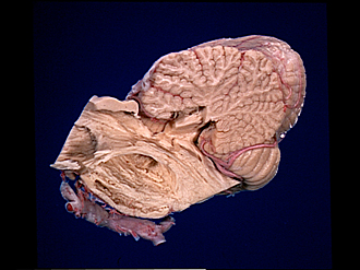
Exploration of the cerebellum and brain stem from the medial aspect
Lateral lemniscus; medial lemniscus; course of trigeminal nerve through pons
Image #27-3
KEYWORDS: Brain, Cerebellum, Midbrain, Peripheral nervous system, Pons.
Creative Commons
Stanford holds the copyright to the David L. Bassett anatomical images and has assigned Creative Commons license Attribution-Share Alike 4.0 International to all of the images.
For additional information regarding use and permissions, please contact the Medical History Center.
Exploration of the cerebellum and brain stem from the medial aspect
Lateral lemniscus; medial lemniscus; course of trigeminal nerve through pons
The brachium conjunctivum has been partly removed and the tegmentum of the mesencephalon further dissected. The pigmented border of the substantia nigra (8) remains, but much of this nucleus was scraped away to demonstrate the cerebral peduncle (7). From the lower border of the substantia nigra white fibrous tissue appears to continue into the tegmentum of the pons.
- Central lobule
- Superior colliculus
- Inferior colliculus
- Lateral lemniscus
- Ala central lobule
- Medial lemniscus
- Cerebral peduncle
- Substantia nigra
- Fibers passing into tegmentum from substantia nigra
- Position of main sensory nucleus of trigeminal nerve and root of trigeminal nerve passing through brachium pontis (middle cerebellar peduncle) (lower pointer)
- Oculomotor nerve (III)
- Superior cerebellar artery
- Posterior cerebral artery right
- Branch of basilar artery to pons
- Superior semilunar lobe (crus I ansiform lobule)
- Horizontal cerebellar sulcus
- Posterior inferior cerebellar artery (PICA)
- Pyramid (vermis)
- Brachium conjunctivum (superior cerebellar peduncle)
- Fourth ventricle
- Nucleus of vestibular nerve
- Facial colliculus (dissected)
- Striae medullares
- Medial longitudinal fasciculus
- Nucleus hypoglossal nerve
- Longitudinal fasciculus (pyramidal) pontine
- Superficial pontine fibers
- Vertebral arteries


