
Bassett Collection of Stereoscopic Images of Human Anatomy
The Posterior inferior aspect of the cerebellum and brain stem
Choroidal branches of posterior inferior cerebellar artery and choroid plexus of fourth ventricle
Image #25-6
KEYWORDS: Brain, Vasculature, Ventricules.
Creative Commons
Stanford holds the copyright to the David L. Bassett anatomical images and has assigned Creative Commons license Attribution-Share Alike 4.0 International to all of the images.

For additional information regarding use and permissions, please contact the Medical History Center.

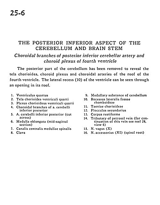
The Posterior inferior aspect of the cerebellum and brain stem
Choroidal branches of posterior inferior cerebellar artery and choroid plexus of fourth ventricle
The posterior part of the cerebellum has been removed to reveal the tela chorioidea, choroid plexus and choroidal arteries of the roof of the fourth ventricle. The lateral recess (10) of the ventricle can be seen through an opening in its roof.
- Fourth ventricle
- Tela chorioidea fourth ventricle
- Choroid plexus fourth ventricle
- Choroidal branches of posterior inferior cerebellar artery
- Posterior inferior cerebellar artery (cut across)
- Medulla oblongata (mid-sagittal section)
- Central canal spinal cord
- Clava
- Medullary substance of cerebellum
- Lateral recess of rhomboid fossa
- Taeniae chorioideae
- Secondary flocculus
- Restiform body (inferior cerebellar peduncle)
- Tributary of petrosal vein (for continuation of this vein see reel 28-6
- Vagus nerve (X)
- Accessory nerve (XI) (spinal root)