Bassett Collection of Stereoscopic Images of Human Anatomy
The Posterior inferior aspect of the cerebellum and brain stem
Foramen of Magendie
Image #25-4
KEYWORDS: Brain, Vasculature, Ventricules.
Creative Commons
Stanford holds the copyright to the David L. Bassett anatomical images and has assigned Creative Commons license Attribution-Share Alike 4.0 International to all of the images.

For additional information regarding use and permissions, please contact the Medical History Center.

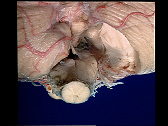
The Posterior inferior aspect of the cerebellum and brain stem
Foramen of Magendie
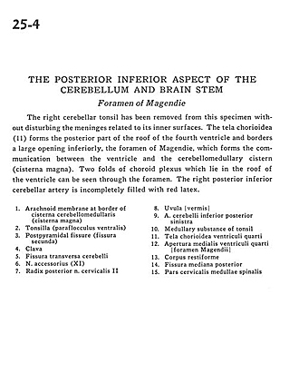
The right cerebellar tonsil has been removed from this specimen without disturbing the meninges related to its inner surfaces. The tela chorioidea (11) forms the posterior part of the roof of the fourth ventricle and borders a large opening inferiorly, the foramen of Magendie, which forms the communication between the ventricle and the cerebellomedullary cistern (cisterna magna). Two folds of choroid plexus which lie in the roof of the ventricle can be seen through the foramen. The right posterior inferior cerebellar artery is incompletely filled with red latex.
- Arachnoid membrane at border of cerebellomedullary cistern (cisterna magna)
- Tonsil (ventral paraflocculus)
- Postpyramidal fissure (fissura secunda)
- Clava
- Transverse cerebellar fissure
- Accessory nerve (CN XI)
- Dorsal root cervical nerve II
- Uvula (vermis)
- Posterior inferior cerebellar artery left
- Medullary substance of tonsil
- Tela chorioidea fourth ventricle
- Medial aperture fourth ventricle foramen of Magendie
- Restiform body (inferior cerebellar peduncle)
- Dorsal median sulcus
- Cervical part of spinal cord