Bassett Collection of Stereoscopic Images of Human Anatomy
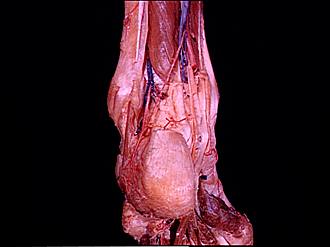
Posterior aspect of left ankle and foot
Peroneus accessorius muscle; long flexor tendons in relation to ankle, posterior view
Image #199-4
KEYWORDS: Ankle, Foot and toes, Muscles and tendons.
Creative Commons
Stanford holds the copyright to the David L. Bassett anatomical images and has assigned Creative Commons license Attribution-Share Alike 4.0 International to all of the images.

For additional information regarding use and permissions, please contact the Medical History Center.


Posterior aspect of left ankle and foot
Peroneus accessorius muscle; long flexor tendons in relation to ankle, posterior view
The peroneus longus and brevis muscles have been cut off. A peroneus accessorius muscle (7) is present with a small muscular portion attached to the lateral side of the calcaneus and a slender tendon extending upward to blend with the posterior intermuscular septum of the leg. The calcaneal tendon has been divided slightly above its attachment of the calcaneus.
- Fibula
- Posterior intermuscular septum
- Tendon of accessory peroneus muscle
- Superior peroneal retinaculum (divided)
- Groove for peroneal tendons
- Calcaneal branch of peroneal artery
- Accessory peroneus muscle
- Tendo calcaneus
- Rete calcaneum
- Peroneus longus muscle (tendon, cut off)
- Tuberosity of calcaneus
- Flexor hallucis longus muscle
- Posterior tibial artery
- Calcaneal branch of the posterior tibial artery
- Tibial nerve (pointer on medial plantar nerve, cut off)
- Lateral plantar nerve
- Flexor retinaculum (opened to expose tendons)
- Flexor hallucis longus muscle (tendon)
- Flexor digitorum longus muscle (tendon)
- Tibialis posterior muscle (tendon)
- Flexor hallucis brevis muscle