Bassett Collection of Stereoscopic Images of Human Anatomy
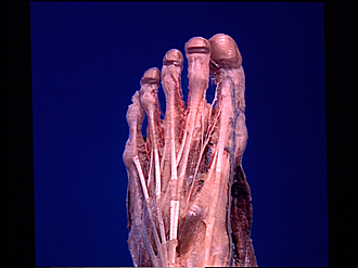
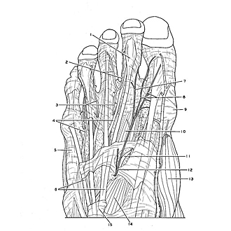
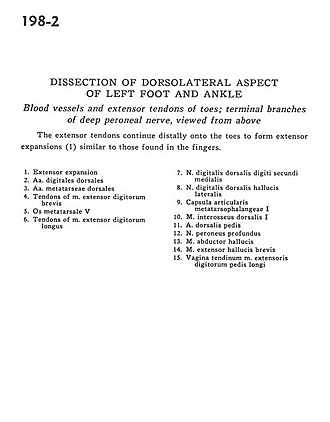
Dissection of dorsolateral aspect of left foot and ankle
Blood vessels and extensor tendons of toes; terminal branches of deep peroneal nerve, viewed from above
Image #198-2
KEYWORDS: Ankle, Foot and toes, Muscles and tendons, Peripheral nervous system, Vasculature.
Creative Commons
Stanford holds the copyright to the David L. Bassett anatomical images and has assigned Creative Commons license Attribution-Share Alike 4.0 International to all of the images.

For additional information regarding use and permissions, please contact the Medical History Center.
Dissection of dorsolateral aspect of left foot and ankle
Blood vessels and extensor tendons of toes; terminal branches of deep peroneal nerve, viewed from above
The extensor tendons continue distally onto the toes to form extensor expansions (1) similar to those found in the fingers.
- Extensor expansion
- Dorsal digital arteries
- Dorsal metatarsal arteries
- Tendons of extensor digitorum brevis muscle
- 5th metatarsal bone
- Tendons of extensor digitorum longus muscle
- Dorsal digital nerve to medial second toe
- Dorsal digital nerve to lateral first toe
- Capsule of 1st metatarsophalangeal joint
- 1st dorsal interosseus muscle
- Dorsalis pedis artery
- Deep peroneal nerve
- Abductor hallucis muscle
- Extensor hallucis brevis muscle
- Sheath of extensor digitorum longus tendon