Bassett Collection of Stereoscopic Images of Human Anatomy
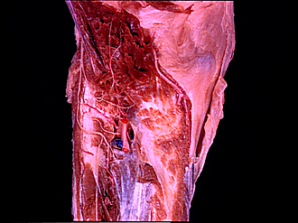
Dissection of posterior aspect of left leg
Nerve supply to popliteus muscle
Image #196-2
KEYWORDS: Leg, Muscles and tendons, Peripheral nervous system.
Creative Commons
Stanford holds the copyright to the David L. Bassett anatomical images and has assigned Creative Commons license Attribution-Share Alike 4.0 International to all of the images.

For additional information regarding use and permissions, please contact the Medical History Center.

Dissection of posterior aspect of left leg
Nerve supply to popliteus muscle
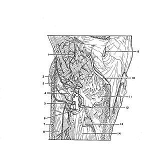
The popliteus has been detached from its insertion on the tibia and reflected upward to display its nerve supply derived from the tibial nerve. The posterior tibial recurrent artery (4) that ramifies within the muscle is a branch of the first part of the anterior tibial artery.
- Popliteus muscle (reflected upward)
- Muscular branch of tibial nerve (to popliteus muscle)
- Muscular branch of tibial nerve (to tibialis posterior muscle)
- Upper pointer: Posterior recurrent tibial artery Lower pointer: Fibular circumflex branch of posterior tibial artery (in this instance it arises from first part of anterior tibial artery)
- Anterior tibial artery
- Posterior tibial artery
- Soleus muscle
- Flexor hallucis longus muscle
- Semimembranosus muscle (insertion)
- Medial inferior genicular artery
- Popliteus muscle (area of insertion)
- Upper pointer: Nutrient artery of tibia Lower pointer: Tendinous arch of tibialis posterior muscle
- Soleus muscle (origin from Soleal line)
- Tibialis posterior muscle