Bassett Collection of Stereoscopic Images of Human Anatomy
Dissection of posterior aspect of left leg
Muscles of deep posterior compartment of leg
Image #195-4
KEYWORDS: Leg, Muscles and tendons.
Creative Commons
Stanford holds the copyright to the David L. Bassett anatomical images and has assigned Creative Commons license Attribution-Share Alike 4.0 International to all of the images.

For additional information regarding use and permissions, please contact the Medical History Center.
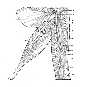
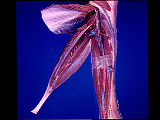
Dissection of posterior aspect of left leg
Muscles of deep posterior compartment of leg
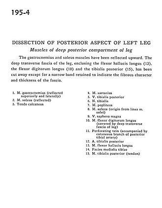
The gastrocnemius and soleus muscles have been reflected upward. The deep transverse fascia of the leg, enclosing the flexor hallucis longus (13), the flexor digitorum longus (10) and the tibialis posterior (15), has been cut away except for a narrow band retained to indicate the fibrous character and thickness of the fascia.
- Gastrocnemius muscle (reflected superiorly and laterally)
- Soleus muscle (reflected)
- Tendo calcaneus
- Sartorius muscle
- Posterior tibial vein
- Tibial nerve
- Popliteus muscle
- Soleus muscle (origin from soleal line)
- Greater saphenous vein
- Flexor digitorum longus muscle (covered by deep transverse fascia of leg)
- Perforating vein (accompanied by cutaneous branch of posterior tibial artery)
- Posterior tibial artery
- Flexor hallucis longus muscle
- Medial surface of tibia
- Tibialis posterior muscle (tendon)