Bassett Collection of Stereoscopic Images of Human Anatomy
Dissection of anterior and medial aspects of thigh
Superficial structures overlying left femoral triangle, anterior view
Image #185-4
KEYWORDS: Fascia, Muscles and tendons, Thigh.
Creative Commons
Stanford holds the copyright to the David L. Bassett anatomical images and has assigned Creative Commons license Attribution-Share Alike 4.0 International to all of the images.

For additional information regarding use and permissions, please contact the Medical History Center.
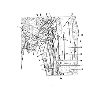
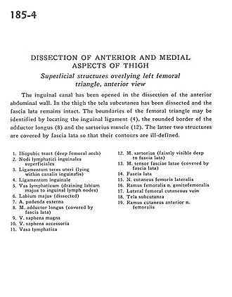
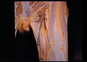
Dissection of anterior and medial aspects of thigh
Superficial structures overlying left femoral triangle, anterior view
The inguinal canal has been opened in the dissection of the anterior abdominal wall. In the thigh the tela subcutanea has been dissected and the fascia lata remains intact. The boundaries of the femoral triangle may be identified by locating the inguinal ligament (4), the rounded border of the adductor longus (8) and the sartorius muscle (12). The latter two structures are covered by fascia lata so that their contours are ill-defined.
- Iliopubic tract (deep femoral arch)
- Superficial inguinal lymph nodes
- Round ligament of uterus (lying within inguinal canal)
- Inguinal ligament
- Lymph duct (draining labium majus to inguinal lymph nodes)
- Labium majus (dissected)
- External pudendal artery
- Adductor longus muscle (covered by fascia lata)
- Greater saphenous vein
- Accessory saphenous vein
- Lymph duct
- Sartorius muscle (faintly visible deep to fascia lata)
- Tensor fasciae latae muscle (covered by fascia lata)
- Fascia lata
- Lateral femoral cutaneous nerve
- Femoral branch of genitofemoral nerve
- Lateral femoral cutaneous vein
- Tela subcutanea
- Cutaneous branch of anterior femoral nerve,