
Bassett Collection of Stereoscopic Images of Human Anatomy
Exploration of the brain from the medial aspect
Relation of stria terminalis to amygdaloid nucleus
Image #18-5
KEYWORDS: Brain, Telencephalon.
Creative Commons
Stanford holds the copyright to the David L. Bassett anatomical images and has assigned Creative Commons license Attribution-Share Alike 4.0 International to all of the images.

For additional information regarding use and permissions, please contact the Medical History Center.
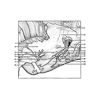

Exploration of the brain from the medial aspect
Relation of stria terminalis to amygdaloid nucleus
The area beneath and behind the posterior limb of the anterior commissure has been dissected away to reveal the extension of the stria terminalis in the region of the amygdaloid nucleus. The complex relations of fibers and gray matter in this area, known as the substantia innominata, do not lend themselves to display by gross dissection. However, a system of fibers (3) is seen passing laterally toward the temporal lobe in this area beneath the lentiform nucleus. These fibers approach the region from the upper surface (stratum zonale) of the thalamus and follow an arching course roughly parallel to the stria terminalis. They have been described as the thalamotemporal tract.
- Frontal part internal capsule
- External capsule
- Fibers entering the "substantia innominata" from the stratum zonale thalami (the thalamotemporal fasciculus)
- Lateral striate arteries
- Anterior commissure
- Basal vein
- Cut surface of frontal lobe
- Middle cerebral artery
- Superior longitudinal fasciculus
- Corona radiata (cut through)
- Lateral ventricle and tapetum (cut across)
- Occipital part radiations corpus callosum
- Sublenticular part of internal capsule (geniculocalcarine tract)
- Fornix (ems)
- Stria terminalis and choroidal fissure
- Hippocampal fissure
- Choroidal branch of posterior cerebral artery
- Hippocampal gyrus
- Uncus hippocampal gyrus
- Choroidal artery (anterior)