
Bassett Collection of Stereoscopic Images of Human Anatomy
Male external genitalia and perineum
Deep perineal space (continued).
Image #168-5
KEYWORDS: Muscles and tendons, Vasculature.
Creative Commons
Stanford holds the copyright to the David L. Bassett anatomical images and has assigned Creative Commons license Attribution-Share Alike 4.0 International to all of the images.

For additional information regarding use and permissions, please contact the Medical History Center.

Male external genitalia and perineum
Deep perineal space (continued).
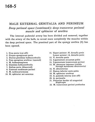
The internal pudendal artery has been divided and removed, together with the artery of the bulb, to reveal more completely the muscles within the deep perineal space. The proximal part of the spongy urethra (4) has been opened.
- Crus of penis (cut off)
- Corpus spongiosum of penis
- Duct of bulbourethral gland
- Spongy part of urethra (opened)
- Bulbospongiosus muscle
- Bulbourethral gland
- Bulb of penis (dissected)
- Central tendon of perineum
- Ischiocavernosus muscle
- External anal sphincter muscle
- Upper pointer: Dorsal nerve of the penis Lower pointer: Dorsal artery of penis
- Dorsal vein of the penis
- Pubic arcuate ligament
- Transverse perineal ligament
- Obturator internus muscle (visible through obturator membrane)
- Obturator nerve
- Inferior pubic ramus
- Sphincter muscle of urethra
- Internal pudendal artery (cut off)
- Perineal artery
- Posterior border of urogenital diaphragm
- Deep transverse perineal muscle