Bassett Collection of Stereoscopic Images of Human Anatomy
Male external genitalia and perineum
Nerve supply to bulbospongiosus muscle
Image #166-7
KEYWORDS: Muscles and tendons, Peripheral nervous system.
Creative Commons
Stanford holds the copyright to the David L. Bassett anatomical images and has assigned Creative Commons license Attribution-Share Alike 4.0 International to all of the images.

For additional information regarding use and permissions, please contact the Medical History Center.
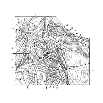
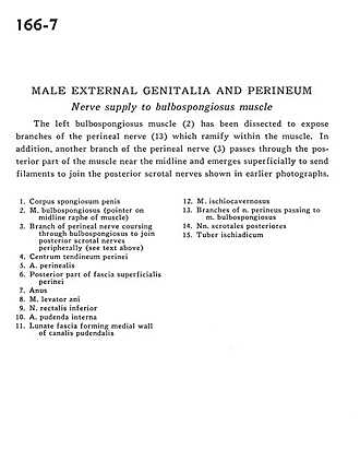
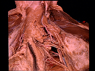
Male external genitalia and perineum
Nerve supply to bulbospongiosus muscle
The left bulbospongiosus muscle (2) has been dissected to expose branches of the perineal nerve (13) which ramify within the muscle. In addition, another branch of the perineal nerve (3) passes through the posterior part of the muscle near the midline and emerges superficially to send filaments to join the posterior scrotal nerves shown in earlier photographs.
- Corpus spongiosum of penis
- Bulbospongiosus muscle (pointer on midline raphe of muscle)
- Branch of perineal nerve coursing through bulbospongiosus to join posterior scrotal nerves peripherally (see text above)
- Central tendon of perineum
- Perineal artery
- Posterior part of superficial perineal fascia
- Anus
- Levator ani muscle
- Inferior rectal nerve
- Internal pudendal artery
- Lunate fascia forming medial wall of pudendal canal
- Ischiocavernosus muscle
- Branches of perineal nerve passing to bulbospongiosus muscle
- Posterior scrotal nerves
- Ischial tuberosity