Bassett Collection of Stereoscopic Images of Human Anatomy
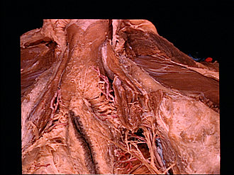
Male external genitalia and perineum
Nerve supply to ischiocavernosus muscle
Image #166-6
KEYWORDS: Muscles and tendons, Perineum, Peripheral nervous system, Vasculature.
Creative Commons
Stanford holds the copyright to the David L. Bassett anatomical images and has assigned Creative Commons license Attribution-Share Alike 4.0 International to all of the images.

For additional information regarding use and permissions, please contact the Medical History Center.

Male external genitalia and perineum
Nerve supply to ischiocavernosus muscle
The ischiocavernosus muscle has been divided posteriorly and spread apart to expose branches of the perineal nerve that supply the muscle. One of these branches terminates in the posterior part of the muscle whereas another branch passes forward to the anterior part of the muscle belly. Posterior scrotal branches of the perineal nerve have been displaced laterally and medially to display the motor branch (21).
- Deep perineal fascia (Buck's fascia)
- Bulbospongiosus muscle
- Ramus of ischium (area indicated by lower pointer covered by periosteum)
- Crus of penis
- Ischiocavernosus muscle
- Perineal artery
- Transverse perineal artery
- Posterior scrotal nerve
- Posterior part of superficial perineal fascia
- Lunate fascia within ischiorectal fossa
- External anal sphincter muscle
- Branches of perineal nerve (spread to reveal nerve to Ischiocavernosus muscle)
- Levator ani muscle
- Corpus spongiosum of penis
- Superficial perineal fascia (reflected)
- Crus of penis
- Nerve to anterior part of Ischiocavernosus muscle
- Ischiocavernosus muscle (dissected)
- Ramus of ischium
- Nerve to posterior part of Ischiocavernosus muscle
- Perineal nerve (branch to ischiocavernosus)
- Internal pudendal artery and vein