Bassett Collection of Stereoscopic Images of Human Anatomy
Creative Commons
Stanford holds the copyright to the David L. Bassett anatomical images and has assigned Creative Commons license Attribution-Share Alike 4.0 International to all of the images.

For additional information regarding use and permissions, please contact the Medical History Center.
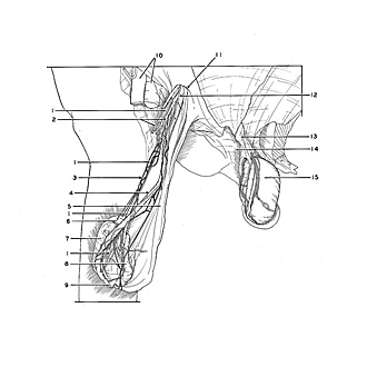
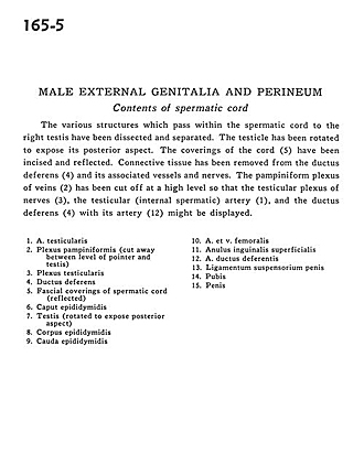
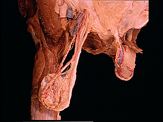
Male external genitalia and perineum
Contents of spermatic cord
The various structures which pass within the spermatic cord to the right testis have been dissected and separated. The testicle has been rotated to expose its posterior aspect. The coverings of the cord (5) have been incised and reflected. Connective tissue has been removed from the ductus deferens (4) and its associated vessels and nerves. The pampiniform plexus of veins (2) has been cut off at a high level so that the testicular plexus of nerves (3), the testicular (internal spermatic) artery (1), and the ductus deferens (4) with its artery (12) might be displayed.
- Testicular artery
- Pampiniform plexus (cut away between level of pointer and testis)
- Testicular plexus
- Ductus deferens
- Fascial coverings of spermatic cord (reflected)
- Head of epididymis
- Testis (rotated to expose posterior aspect)
- Body of epididymis
- Tail of epididymis
- Femoral artery and vein
- Superficial inguinal ring
- Artery of ductus deferens
- Suspensory ligament of the penis
- Pubis
- Glans penis