Bassett Collection of Stereoscopic Images of Human Anatomy
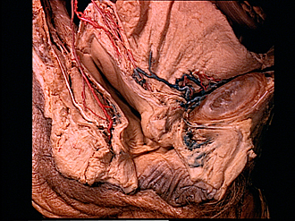
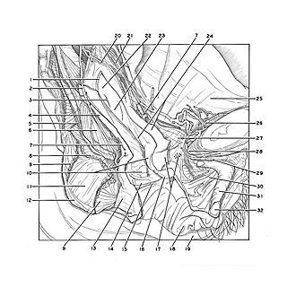
Dissection of female pelvis from a lateral approach
Interior of vagina, viewed from right side
Image #161-1
KEYWORDS: Vagina, Muscles and tendons, Ovary.
Creative Commons
Stanford holds the copyright to the David L. Bassett anatomical images and has assigned Creative Commons license Attribution-Share Alike 4.0 International to all of the images.

For additional information regarding use and permissions, please contact the Medical History Center.
Dissection of female pelvis from a lateral approach
Interior of vagina, viewed from right side
The vagina has been opened along its right border. Its walls have been separated to permit a view of the interior.
- Fornix of vagina
- Middle rectal artery
- Opening of uterus
- Posterior wall of vagina
- Superior fascia of pelvic diaphragm
- Rectum
- Vaginal venous plexus
- Levator ani muscle (cut off)
- External anal sphincter muscle (superficial and deep portions, partially removed)
- Upper pointer: Urethral carina Lower pointer: Posterior neck fold
- Muscular tunic of anal canal (longitudinal layer)
- External anal sphincter muscle (subcutaneous part)
- Central tendon of perineum
- Hymenal caruncles
- Vestibule of vagina
- External urethral opening
- Urogenital diaphragm
- Labium minus
- Labium majus
- Vaginal artery
- Inferior vesical artery
- Posterior labium uterine opening
- Anterior labium uterine opening
- Left pointer: Posterior vaginal wall anterior Right pointer: Anterior wall of vagina
- Urinary bladder
- Vesical venous plexus
- Vesical sphincter muscle
- Pubic arcuate ligament (cut in midline)
- Dorsal vein of clitoris
- Commissure of vestibular bulb
- Body of clitoris
- Glans of clitoris