Bassett Collection of Stereoscopic Images of Human Anatomy
Dissection of female pelvis froma lateral approach
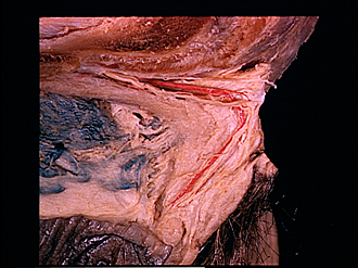
Close-up view of clitoris
Image #159-3
KEYWORDS: Muscles and tendons.
Creative Commons
Stanford holds the copyright to the David L. Bassett anatomical images and has assigned Creative Commons license Attribution-Share Alike 4.0 International to all of the images.
For additional information regarding use and permissions, please contact the Medical History Center.
Dissection of female pelvis froma lateral approach
Close-up view of clitoris
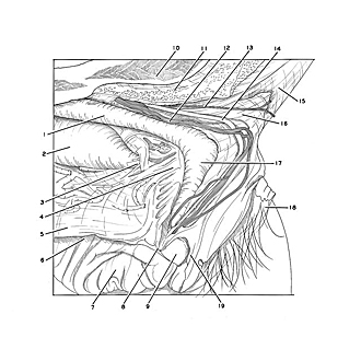
The structure and relations of the clitoris are shown in this close-up view of the specimen illustrated in the previous photograph. The ischiocavernosus and bulbospongiosus muscles have been removed from the dissection. The right crus, body and glans of the clitoris are exposed. A fibrous commissural extension (4) from the pars intermedia of the vestibular bulb, joined by its fellow of the opposite side, passes forward to fuse with the clitoris.
- Crus of clitoris
- Vestibular bulb (cavernous spaces within erectile tissue filled with blue latex)
- Intermediate part of bulb of vestibule
- Commissure of vestibular bulb (joined anteriorly with body of clitoris)
- Labium minus (dissected)
- Vestibule of vagina
- Labium minus
- Frenulum of clitoris
- Glans of clitoris
- Pubic bone (partially resected)
- Tendon of origin of gracilis muscle (cut off)
- Dorsal nerve of clitoris
- Cutaneous branch of dorsal nerve of clitoris
- Dorsal artery of clitoris
- Pubic symphysis
- Suspensory ligament of clitoris
- Body of clitoris
- Skin of mons pubis
- Prepuce of clitoris


