Bassett Collection of Stereoscopic Images of Human Anatomy
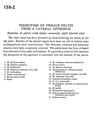
Dissection of female pelvis from a lateral approach
Exterior of pelvis with femur removed, right lateral view
Image #158-2
KEYWORDS: Bones joints cartilage, Muscles and tendons, Vasculature.
Creative Commons
Stanford holds the copyright to the David L. Bassett anatomical images and has assigned Creative Commons license Attribution-Share Alike 4.0 International to all of the images.

For additional information regarding use and permissions, please contact the Medical History Center.
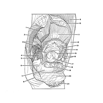
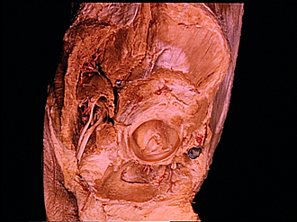
Dissection of female pelvis from a lateral approach
Exterior of pelvis with femur removed, right lateral view
The right thigh has been detached by disarticulating the femur at the hip joint. Muscles of the gluteal region have been cut off in various ways to demonstrate their interrelations. The obturator externus and pectineus muscles have been completely removed. The periosteum has been stripped from the rami of the pubis and ischium. In succeeding views of this sequence the dissection of the specimen is continued into the interior of the pelvis.
- Gluteus medius muscle
- Gluteus maximus muscle
- Piriform muscle
- Obturator internus muscle (cut off)
- Sciatic nerve
- Ischial tuberosity
- Ramus of ischium
- Anus
- External oblique muscle
- Iliac crest
- Gluteus minimus muscle
- Aponeurosis External oblique muscle
- Rectus femoris muscle (origin, cut off)
- Iliopsoas muscle (cut off)
- Joint capsule of coccyx
- Femoral artery and vein
- Acetabulum (pointer on ligamentum teres)
- Pubic bone
- Obturator membrane
- Superficial perineal fascia (membranous layer)
- Labium majus