Bassett Collection of Stereoscopic Images of Human Anatomy
Ligaments and joints of lumbosacral spine and pelvic girdle
Sacroiliac joint opened; interosseous sacroiliac ligament, left lateral view
Image #156-2
KEYWORDS: Central nervous system, Bones joints cartilage, Vasculature.
Creative Commons
Stanford holds the copyright to the David L. Bassett anatomical images and has assigned Creative Commons license Attribution-Share Alike 4.0 International to all of the images.

For additional information regarding use and permissions, please contact the Medical History Center.
Ligaments and joints of lumbosacral spine and pelvic girdle
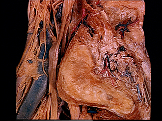
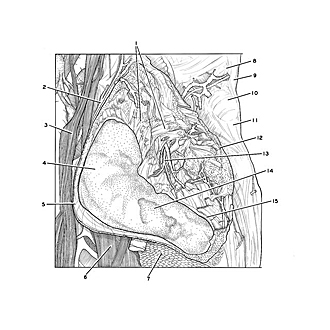
Sacroiliac joint opened; interosseous sacroiliac ligament, left lateral view
The left ilium has been detached to display the auricular surface of the sacrum and the interosseous sacroiliac ligaments.
- Interosseous sacroiliac ligament (pointer indicates only a small part of the area occupied by the numerous bands that comprise these ligaments which have been cut at their attachments to the ilium)
- Iliolumbar artery
- Lumbosacral trunk
- Articular surface of sacrum
- Ventral sacroiliac ligament
- Sacral nerve I
- Piriform muscle (cut across)
- Spinous process vertebra L. V
- Supraspinous ligament
- Interspinous ligament L. V - S. I (convex in shape due to compression by extension of vertebra on sacrum)
- Middle sacral crest
- Dorsal sacroiliac ligament (cut across)
- Iliac branch of iliolumbar artery
- Area of articular surface in which fibrocartilage has been damaged (remainder of auricular surface intact)
- Joint capsule (lined internally by synovial membrane)