
Bassett Collection of Stereoscopic Images of Human Anatomy
Exploration of liver, gall bladder, pancreas, duodenum and spleen
General view of organs in situ, stomach opened
Image #145-6
KEYWORDS: Gallbladder, Liver, Pancreas, Spleen, Stomach, Overview.
Creative Commons
Stanford holds the copyright to the David L. Bassett anatomical images and has assigned Creative Commons license Attribution-Share Alike 4.0 International to all of the images.

For additional information regarding use and permissions, please contact the Medical History Center.

Exploration of liver, gall bladder, pancreas, duodenum and spleen
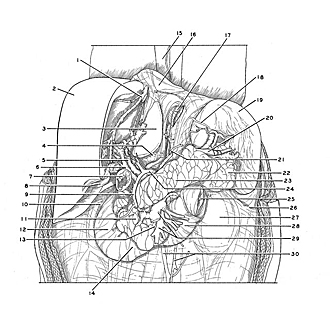
General view of organs in situ, stomach opened
The stomach has been removed by dividing the esophagus (17) above the cardia and by severing the duodenum (7) close to the pylorus.
- Hepatic vein
- Liver
- Caudate lobe
- Vestibule of omental bursa
- Portal vein
- Common bile duct
- Superior part of duodenum (cut across)
- Gallbladder
- Gastroduodenal artery
- Prominence of right kidney (in background)
- Head of pancreas
- Descending part of duodenum
- Uncinate process of pancreas
- Horizontal part of duodenum
- Esophagus
- Diaphragm
- Abdominal part of esophagus
- Line of reflection of peritoneum onto spleen from gastrosplenic ligament
- Spleen
- Splenic artery (accompanied by vein and nerves)
- Left gastric artery
- Tail of pancreas
- Body of pancreas
- Common hepatic artery
- Jejunum (cut across)
- Line of attachment of descending colon (pointer on cut edge of peritoneum)
- Left kidney (covered by fascia)
- Ascending part of duodenum
- Superior mesenteric artery and vein
- Inferior mesenteric artery and vein