Bassett Collection of Stereoscopic Images of Human Anatomy
Dissection of male inguinal region and spermatic cord
Internal aspect of deep inguinal ring and related structures of right side (continued)
Image #136-7
KEYWORDS: Overview.
Creative Commons
Stanford holds the copyright to the David L. Bassett anatomical images and has assigned Creative Commons license Attribution-Share Alike 4.0 International to all of the images.

For additional information regarding use and permissions, please contact the Medical History Center.
Dissection of male inguinal region and spermatic cord
Internal aspect of deep inguinal ring and related structures of right side (continued)
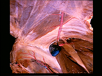
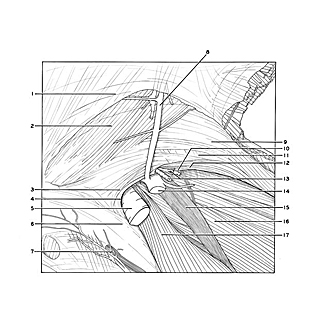
The transversalis and ilias fascias have been cut away from the specimen which was shown in the previous view. The ductus deferens (13) is visible passing into the upper part of the inguinal canal. A small muscle fasciculus (12) which originates with the transversus abdominis passes into the inguinal canal to form a portion of the cremaster muscle. The lacunar ligament (4) passes slightly lateral to the margin of the falx inguinalis (3) and thus forms the medial boundary of the vascular compartment deep to the inguinal ligament.
- Transversalis fascia (partially cut away)
- Rectus abdominis muscle
- Conjoint tendon
- Lacunar ligament
- External iliac vein (displaced somewhat medially)
- Pectineal ligament (Cooper's ligament)
- Obturator canal
- Inferior epigastric artery
- Transversus abdominis muscle (pointer on aponeurotic fibers, note other parallel fibers at lower level and muscle fibers continuing laterally)
- Inguinal ligament (white band in depths of dissected wall it has been elevated)
- Internal oblique muscle (visible through aponeurosis of transversus abdominis)
- Cremaster muscle (fibers originating with transversus abdominis muscle)
- Upper pointer: Testicular artery Lower pointer: Ductus deferens (passing into inguinal canal)
- External iliac artery (elevated)
- Femoral nerve
- Iliacus muscle
- Psoas major muscle