Bassett Collection of Stereoscopic Images of Human Anatomy
Detailed dissection of heart
Interior of left ventricle.
Image #122-6
KEYWORDS: Heart, Left heart.
Creative Commons
Stanford holds the copyright to the David L. Bassett anatomical images and has assigned Creative Commons license Attribution-Share Alike 4.0 International to all of the images.

For additional information regarding use and permissions, please contact the Medical History Center.
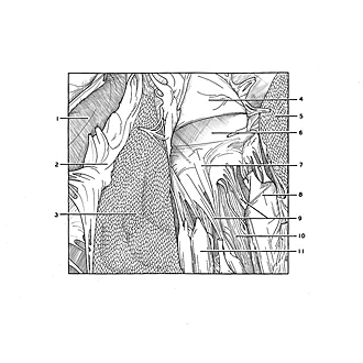
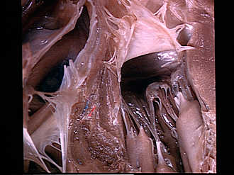
Detailed dissection of heart
Interior of left ventricle.
The anterior cusp of the mitral valve has been freed of its chordal attachments and retracted upward. By this means the posterior cusp of the valve (7), the valvular ostium (6) and the interior of the left atrium have been brought to view.
- Right atrioventricular opening
- Septal (medial) cusp of tricuspid valve
- Muscular part interventricular septum
- Anterior cusp of mitral valve (retracted)
- Cut edge of wall of left ventricle
- Left atrioventricular opening (left atrium visible in background)
- Posterior cusp of mitral valve
- Anterior papillary muscle
- Chordae tendineae
- Left ventricle
- Posterior papillary muscle