Bassett Collection of Stereoscopic Images of Human Anatomy
Detailed dissection of heart
Interior of left atrium, posterior view
Image #122-1
KEYWORDS: Heart, Left heart.
Creative Commons
Stanford holds the copyright to the David L. Bassett anatomical images and has assigned Creative Commons license Attribution-Share Alike 4.0 International to all of the images.

For additional information regarding use and permissions, please contact the Medical History Center.
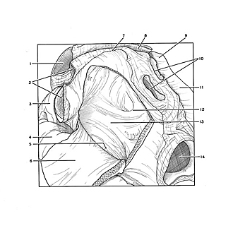
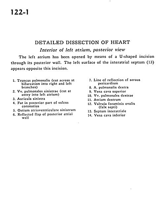
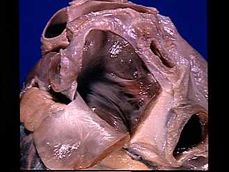
Detailed dissection of heart
Interior of left atrium, posterior view
The left atrium has been opened by means of a U-shaped incision through its posterior wall. The left surface of the interatrial septum (13) appears opposite this incision.
- Pulmonary trunk (cut across at bifurcation into right and left branches)
- Left pulmonary veins (cut at entry into left atrium)
- Left auricle
- Fat in posterior part of coronary sulcus
- Left atrioventricular opening
- Reflected flap of posterior atrial wall
- Line of reflection of serous pericardium
- Right pulmonary artery
- Superior vena cava
- Right pulmonary veins
- Right atrium
- Valve of foramen ovale (falx septi)
- Interatrial septum
- Inferior vena cava