
Bassett Collection of Stereoscopic Images of Human Anatomy
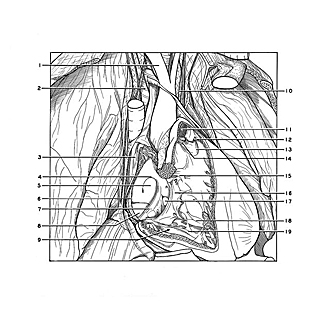
Dissection of pericardium and heart in situ
Interior of right atrium and right ventricle
Image #117-4
KEYWORDS: Heart, Pericardial sac, Right heart.
Creative Commons
Stanford holds the copyright to the David L. Bassett anatomical images and has assigned Creative Commons license Attribution-Share Alike 4.0 International to all of the images.

For additional information regarding use and permissions, please contact the Medical History Center.

Dissection of pericardium and heart in situ
Interior of right atrium and right ventricle
The latex cast has been removed from the specimen shown in the previous view.
- Brachiocephalic trunk
- Trachea
- Sino-atrial node
- Border of fossa ovalis
- Fossa ovalis
- Crista terminalis
- Valve of inferior vena cava
- Latex cast cut across at junction of vena cava inferior with vena caval sinus
- Right ventricle
- Inferior cardiac branch of vagus nerve
- Pulmonary trunk
- Anterior semilunar cusp, (cut across)
- Left semilunar cusp
- Right semilunar cusp (cut across) (11-13 make up the pulmonary valve)
- Supraventricular crest
- Septal (medial) cusp of tricuspid valve
- Latex cast in coronary sinus
- Anterior papillary muscle
- Posterior papillary muscle