Bassett Collection of Stereoscopic Images of Human Anatomy
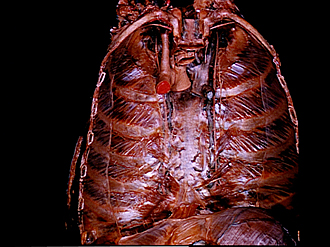
Dissection of breast and anterolateral thoracic wall
Anterior thoracic wall, internal aspect
Image #116-1
KEYWORDS:
Creative Commons
Stanford holds the copyright to the David L. Bassett anatomical images and has assigned Creative Commons license Attribution-Share Alike 4.0 International to all of the images.

For additional information regarding use and permissions, please contact the Medical History Center.
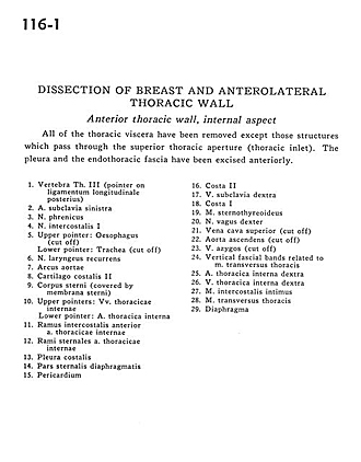
Dissection of breast and anterolateral thoracic wall
Anterior thoracic wall, internal aspect
All of the thoracic viscera have been removed except those structures which pass through the superior thoracic aperture (thoracic inlet). The pleura and the endothoracic fascia have been excised anteriorly.
- Vertebra Th. III (pointer on posterior longitudinal ligament)
- Left subclavian artery
- Phrenic nerve
- Intercostal nerve
- Upper pointer: Esophagus (cut off) Lower pointer: Trachea (cut off)
- Recurrent laryngeal nerve
- Aortic arch
- Costal cartilage II
- Body of sternum (covered by sternal membrane)
- Upper pointers: Internal thoracic veins Lower pointer: Internal thoracic artery
- Anterior intercostal branch internal thoracic artery
- Sternal branches internal thoracic artery
- Costal pleura
- Sternal part of diaphragm
- Pericardium
- Rib II
- Right subclavian vein
- Rib I
- Sternothyroid muscle
- Vagus nerve right
- Superior vena cava (cut off)
- Ascending aorta
- Azygos vein (cut off)
- Vertical fascial bands related to transversus thoracis muscle
- Right internal thoracic artery
- Left internal thoracic vein
- Innermost intercostal muscle
- Transversus thoracis muscle
- Diaphragm