Bassett Collection of Stereoscopic Images of Human Anatomy
Exploration of the brain from its superior aspect
Anterior nucleus of thalamus and mammillothalamic tract; inferior horn of lateral ventricle
Image #11-7
KEYWORDS: Brain, Diencephalon, Telencephalon, Ventricules.
Creative Commons
Stanford holds the copyright to the David L. Bassett anatomical images and has assigned Creative Commons license Attribution-Share Alike 4.0 International to all of the images.
For additional information regarding use and permissions, please contact the Medical History Center.
Exploration of the brain from its superior aspect
Anterior nucleus of thalamus and mammillothalamic tract; inferior horn of lateral ventricle
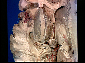
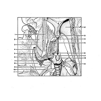
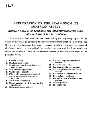
The thalamus has been further dissected by cutting away a part of the anterior nucleus and exposing the mammillothalamic tract in its course into this area. The tapetum has been removed to display the inferior horn of the lateral ventricle, the tail of the caudate nucleus and the downward continuation of those fibers of the stratum zonale of the thalamus seen in the previous view.
- Corona radiata
- Lentiform nucleus
- Inferior occipitofrontal fasciculus (cut across)
- Hippocampal digitations
- Caudate nucleus (tail)
- Fibers of stratum zonale thalami
- Choroid plexus lateral ventricle
- Collateral eminence
- Calcar avis (not well defined in this specimen)
- Bulbus posterior horn
- External sagittal stratum (cut across)
- Head of caudate nucleus
- Stria terminalis (cut across)
- Anterior nucleus of thalamus (cut through)
- Mamillothalamic tract
- Fasciculus retroflexus
- Posterior stalk of thalamus (cut across)
- Pineal body
- Hippocampus


