Bassett Collection of Stereoscopic Images of Human Anatomy
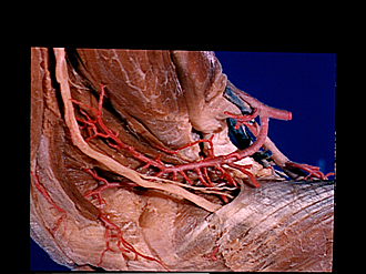
Dorsal aspect of right forearm
Recurrent radial artery
Image #107-5
KEYWORDS: Elbow, Forearm, Peripheral nervous system.
Creative Commons
Stanford holds the copyright to the David L. Bassett anatomical images and has assigned Creative Commons license Attribution-Share Alike 4.0 International to all of the images.

For additional information regarding use and permissions, please contact the Medical History Center.


Dorsal aspect of right forearm
Recurrent radial artery
Several branches of the artery have been cut off. These supplied the extensor muscles shown in previous views.
- Brachialis muscle
- Extensor carpi radialis longus muscle (cut close to origin)
- Deep branch of radial nerve
- Terminal superficial branch of recurrent radial artery
- Upper pointer: Origin of extensor carpi radialis brevis muscle Lower painter: Origin of common extensor digitorum muscle
- Cubital joint capsule (position of head of radius)
- Biceps brachii muscle
- Muscular branch radial
- Radial artery
- Recurrent radial artery
- Ulnar vein
- Ulnar artery
- Supinator muscle (superficial belly)
- Supinator muscle (deep lamina)