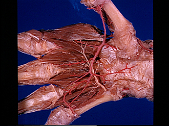
Bassett Collection of Stereoscopic Images of Human Anatomy
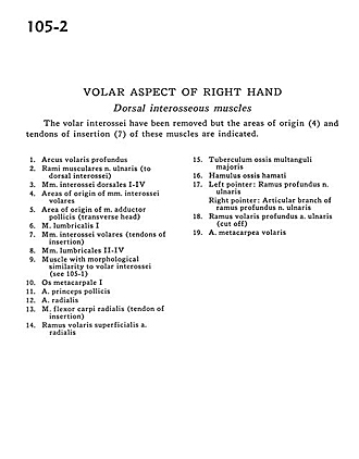
Volar aspect of right hand
Dorsal interosseous muscles
Image #105-2
KEYWORDS: Hand and fingers, Vasculature.
Creative Commons
Stanford holds the copyright to the David L. Bassett anatomical images and has assigned Creative Commons license Attribution-Share Alike 4.0 International to all of the images.

For additional information regarding use and permissions, please contact the Medical History Center.
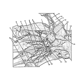
Volar aspect of right hand
Dorsal interosseous muscles
The volar interossei have been removed but the areas of origin (4) and tendons of insertion (7) of these muscles are indicated.
- Deep palmar arch
- Muscular branches of ulnar nerve (to dorsal interossei)
- Dorsal interosseous muscles I-IV
- Areas of origin of anterior interosseous muscles
- Area of origin of adductor pollicis muscle (transverse head)
- Lumbrical muscle I
- Anterior interosseous muscles (tendons of insertion)
- Lumbrical muscles II-IV
- Muscle with morphological similarity to anterior interossei (see 105-3)
- Metacarpal I
- Princeps pollicis artery
- Radial artery
- Flexor carpi radialis muscle (tendon of insertion)
- Superficial anterior branch radial artery
- Tubercle of trapezium bone
- Hamulus of hamate bone
- Left pointer: Deep branch of ulnar nerve Right pointer: Articular branch of deep branch of ulnar nerve
- Deep anterior branch ulnar artery (cut off)
- Anterior metacarpal artery