
Bassett Collection of Stereoscopic Images of Human Anatomy
Volar aspect of right hand
Thenar muscles; deep volar arch; deep branch of ulnar nerve
Image #104-5
KEYWORDS: Fascia ligaments and tendons, Hand and fingers, Neuralnetwork, Vasculature.
Creative Commons
Stanford holds the copyright to the David L. Bassett anatomical images and has assigned Creative Commons license Attribution-Share Alike 4.0 International to all of the images.

For additional information regarding use and permissions, please contact the Medical History Center.
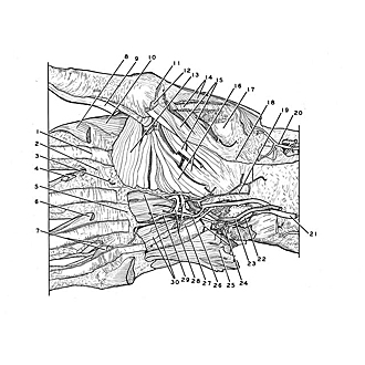
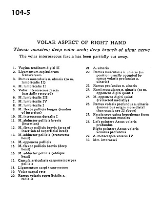
Volar aspect of right hand
Thenar muscles; deep volar arch; deep branch of ulnar nerve
The volar interosseous fascia has been partially cut away.
- Tendon of digital sheath II
- Deep transverse metacarpal ligament
- Muscular branch of ulnar nerve (to lumbrical muscle II)
- Lumbrical muscle II
- Anterior interosseous fascia (partially resected)
- Lumbrical muscle III
- Lumbrical muscle IV
- Lumbrical muscle I
- Flexor pollicis longus muscle (tendon of insertion)
- Dorsal interosseous muscle I
- Abductor pollicis brevis muscle (insertion)
- Flexor pollicis brevis muscle (area of insertion of superficial head)
- Adductor pollicis muscle (transverse head)
- Opponens pollicis muscle
- Flexor pollicis brevis muscle (deep head)
- Adductor pollicis muscle (oblique head)
- Metacarpotrapezial joint capsule
- Transverse carpal ligament
- Volar carpal rete
- Superficial anterior branch radial artery
- Ulnar artery
- Muscular branch ulnar artery (in position usually occupied by deep anterior branch ulnar artery)
- Deep branch of ulnar nerve
- Muscular branches of ulnar nerve (to opponens digiti minimi muscle)
- Opponens digiti minimi muscle (retracted medially)
- Deep anterior branch ulnar artery (anomalous origin more distal than usual see 22 above)
- Fascia separating hypothenar from interosseous muscles
- Left pointer: Deep palmar arch Right pointer: Deep anterior venous arch
- Anterior metacarpal artery IV
- Interosseous muscles