
Bassett Collection of Stereoscopic Images of Human Anatomy
Exploration of the meninges and brain in situ
Cerebellomedullary cistern (cisterna magna)
Image #1-7
KEYWORDS: Brain, Cerebellum, Meninges, Central nervous system.
Creative Commons
Stanford holds the copyright to the David L. Bassett anatomical images and has assigned Creative Commons license Attribution-Share Alike 4.0 International to all of the images.

For additional information regarding use and permissions, please contact the Medical History Center.

Exploration of the meninges and brain in situ
Cerebellomedullary cistern (cisterna magna)
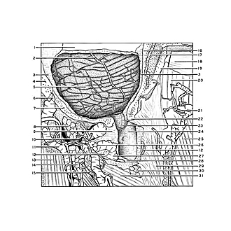
The arachnoid membrane of the posterior cranial fossa and upper part of the vertebral canal is exposed to the left of the midline. The left half of the occipital bone, including a part of the margin of the foramen magnum (22), has been cut away extensively. A large window has been cut in the dura mater covering the posterior cranial fossa and upper cervical spinal cord. As the dura passes through the foramen magnum its outer portion blends with the dense fibrous tissue of the posterior atlanto-occipital membrane (9). A plexus of veins is embedded in this fibrous tissue. The inner layer of dura is continuous with that which covers the spinal cord.
- Transverse sinus (opened)
- Inferior cerebellar vein (lateral posterior cerebellar vein)
- Dura mater
- Mastoid part of temporal bone
- Cerebellum (arachnoid intact over its surface)
- Mastoid emissary
- Occipital bone
- Obliquus capitis superior muscle (cut across)
- Posterior atlanto-occipital membrane
- Suboccipital nerve
- Occipitalis vein
- Posterior arch of atlas (cut across)
- Splenius capitis muscle (reflected laterally)
- Semispinalis capitis muscle (cut across)
- Occipitalis major nerve
- Internal plate of occipital bone
- Occipital diploic vein
- Transverse nuchal muscle (tendinous)
- External plate of occipital bone
- Semispinalis capitis muscle
- Subcutaneous branch of occipital artery
- Occipital bone near margin of foramen magnum
- Cerebellar tonsil lying deep to arachnoid membrane (Note: this extends downward slightly more than usual)
- Arachnoid (overlying cerebellomedulary cistern)
- Trapezius muscle
- Vertebral artery (visible deep to venous plexus which surrounds it in the vertebral artery sulcus)
- Posterior vertebral venous plexus
- Dura mater (cut edge)
- Ligamentum nuchae (cervical muscles removed from its left side)
- Rectus capitis posterior major muscle (cut across near its origin from the spine of the axis)
- Inferior obliquus capitis muscle