Bassett Collection of Stereoscopic Images of Human Anatomy
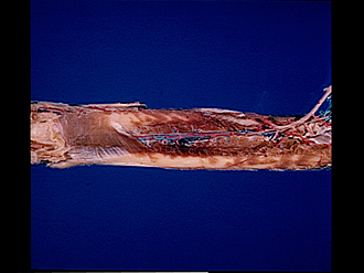
Volar aspect of forearm
Pronator quadratus muscle; origins of deep layer of flexor muscles
Image #99-4
KEYWORDS: Forearm, Vasculature.
Creative Commons
Stanford holds the copyright to the David L. Bassett anatomical images and has assigned Creative Commons license Attribution-Share Alike 4.0 International to all of the images.

For additional information regarding use and permissions, please contact the Medical History Center.
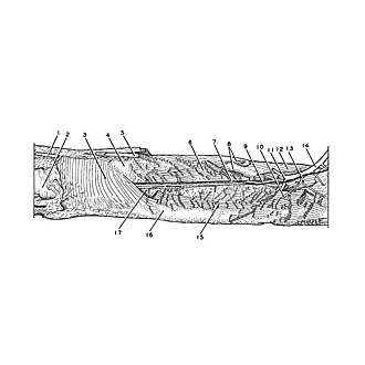
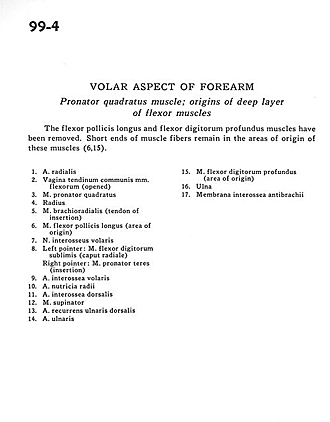
Volar aspect of forearm
Pronator quadratus muscle; origins of deep layer of flexor muscles
The flexor pollicis longus and flexor digitorum profundus muscles have been removed. Short ends of muscle fibres remain in the areas of origin of these muscles (6,15).
- Radial artery
- Sheath of common tendon of flexor muscles (opened)
- Pronator quadratus muscle
- Radius
- Brachioradialis muscle (tendon of insertion)
- Flexor pollicis longus muscle (area of origin)
- Anterior interosseous nerve
- Left pointer: Flexor digitorum superficialis (radial head) Right pointer: Pronator teres muscle (insertion)
- Anterior interosseous artery
- Nutritive artery of radius
- Dorsal interosseous artery
- Supinator muscle
- Dorsal recurrent ulnar artery
- Ulnar artery
- Flexor digitorum profundus muscle (area of origin)
- Ulna
- Antebrachial interosseous membrane