
Bassett Collection of Stereoscopic Images of Human Anatomy
Volar aspect of forearm
Nerve and blood supply to flexor digitorum sublimis muscle
Image #98-3
KEYWORDS: Forearm, Neuralnetwork, Peripheral nervous system, Vasculature.
Creative Commons
Stanford holds the copyright to the David L. Bassett anatomical images and has assigned Creative Commons license Attribution-Share Alike 4.0 International to all of the images.

For additional information regarding use and permissions, please contact the Medical History Center.

Volar aspect of forearm
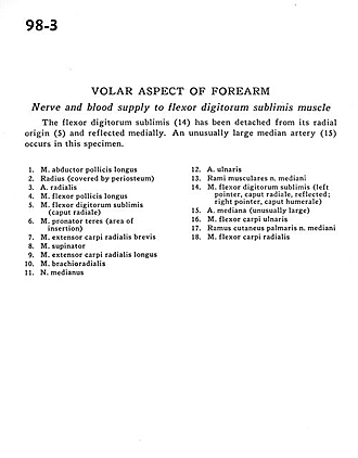
Nerve and blood supply to flexor digitorum sublimis muscle
The flexor digitorum sublimis (14) has been detached from its radial origin (5) and reflected medially. An unusually large median artery (15) occurs in this specimen.
- Abductor pollicis longus muscle
- Radius (covered by periosteum)
- Radial artery
- Flexor pollicis longus muscle
- Flexor digitorum superficialis (radial head)
- Pronator teres muscle (area of insertion)
- Extensor carpi radialis brevis muscle
- Supinator muscle
- Extensor carpi radialis longus muscle
- Brachioradialis muscle
- Median nerve
- Ulnar artery
- Muscular branches of median nerve
- Flexor digitorum superficialis (left pointer, radial head, reflected; right pointer, humeral head)
- Median artery (unusually large)
- Flexor carpi ulnaris muscle
- Palmar cutaneous branch of median nerve
- Flexor carpi radialis muscle