
Bassett Collection of Stereoscopic Images of Human Anatomy
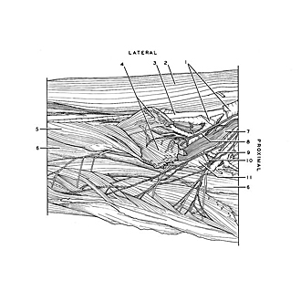
Volar aspect of forearm
Nerve supply to right pronator teres and flexor carpi radialis muscles
Image #97-7
KEYWORDS: Neuralnetwork, Vasculature.
Creative Commons
Stanford holds the copyright to the David L. Bassett anatomical images and has assigned Creative Commons license Attribution-Share Alike 4.0 International to all of the images.

For additional information regarding use and permissions, please contact the Medical History Center.
Volar aspect of forearm
Nerve supply to right pronator teres and flexor carpi radialis muscles
The humeral head (4) of the pronator teres has been divided and its fascicles separated to display branches of the median nerve and ulnar artery within the muscle. The flexor carpi radialis muscle has been retracted medially and dissected in a similar manner.
- Vena comitans of radial artery
- Brachioradialis muscle
- Radial artery
- Pronator teres muscle (humeral head)
- Pronator teres muscle (pointer near insertion on radius)
- Flexor digitorum superficialis
- Median nerve
- Muscular branch of median nerve (to both heads of pronator teres muscle)
- Muscular branch of median nerve (to flexor carpi radialis muscle)
- Anterior recurrent ulnar artery
- Pronator teres muscle (ulnar head)