Bassett Collection of Stereoscopic Images of Human Anatomy
Volar aspect of forearm
Superficial nerves and vessels of right forearm
Image #96-6
KEYWORDS: Fascia ligaments and tendons, Forearm, Neuralnetwork, Peripheral nervous system.
Creative Commons
Stanford holds the copyright to the David L. Bassett anatomical images and has assigned Creative Commons license Attribution-Share Alike 4.0 International to all of the images.

For additional information regarding use and permissions, please contact the Medical History Center.
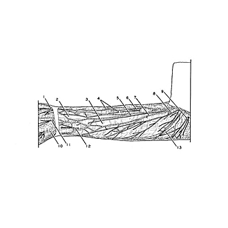
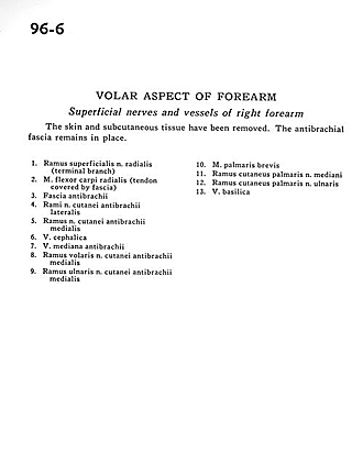
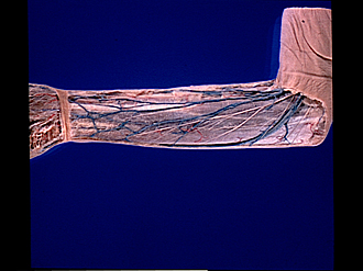
Volar aspect of forearm
Superficial nerves and vessels of right forearm
The skin and subcutaneous tissue have been removed. The antibrachial fascia remains in place.
- Superficial branch of radial nerve (terminal branch)
- Flexor carpi radialis muscle (tendon covered by fascia)
- Antibrachial fascia
- Branches of lateral antebrachial cutaneous nerve
- Branch middle antebrachial cutaneous nerve
- Cephalic vein
- Median antebrachial vein
- Anterior branch middle antebrachial cutaneous nerve
- Ulnar branch of middle antebrachial cutaneous nerve
- Palmaris brevis muscle
- Palmar cutaneous branch of median nerve
- Palmar cutaneous branch of ulnar nerve
- Basilic vein