Bassett Collection of Stereoscopic Images of Human Anatomy
Arm
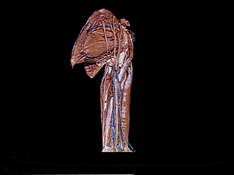
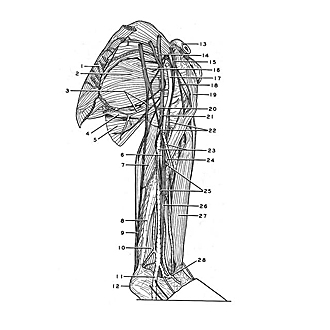
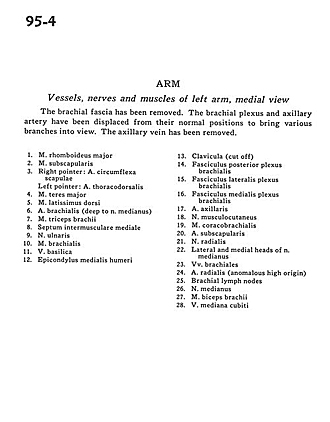
Vessels, nerves and muscles of left arm, medial view
Image #95-4
KEYWORDS: Neuralnetwork, Peripheral nervous system, Upper arm, Vasculature.
Creative Commons
Stanford holds the copyright to the David L. Bassett anatomical images and has assigned Creative Commons license Attribution-Share Alike 4.0 International to all of the images.

For additional information regarding use and permissions, please contact the Medical History Center.
Arm
Vessels, nerves and muscles of left arm, medial view
The brachial fascia has been removed. The brachial plexus and axillary artery have been displayed from their normal positions to bring various branches into view. The axillary vein has been removed.
- Rhomboid major muscle
- Subscapularis muscle
- Right pointer: Circumflex artery of scapula Left pointer: Thoracodorsal artery
- Teres major muscle
- Latissimus dorsi muscle
- Brachial artery (deep to median nerve)
- Triceps brachii muscle
- Medial intermuscular septum
- Ulnar nerve
- Brachialis muscle
- Basilic vein
- Medial epicondyle of humerus
- Clavicle (cut off)
- Posterior cord of brachial plexus
- Lateral cord of brachial plexus
- Middle cord of brachial plexus
- Axillary artery
- Musculocutaneous nerve
- Coracobrachialis muscle
- Subscapular artery
- Radial nerve
- Lateral and medial heads of median nerve
- Brachial veins
- Radial artery (anomalous high origin)
- Brachial lymph nodes
- Median nerve
- Biceps brachii muscle
- Median cubital vein