Bassett Collection of Stereoscopic Images of Human Anatomy
Shoulder
Fascial planes anterior to left shoulder joint
Image #92-4
KEYWORDS: Fascia ligaments and tendons, Muscles and tendons, Shoulder, Vasculature.
Creative Commons
Stanford holds the copyright to the David L. Bassett anatomical images and has assigned Creative Commons license Attribution-Share Alike 4.0 International to all of the images.
For additional information regarding use and permissions, please contact the Medical History Center.
Shoulder
Fascial planes anterior to left shoulder joint
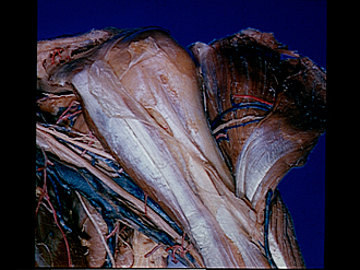
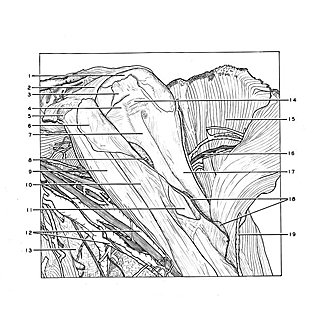
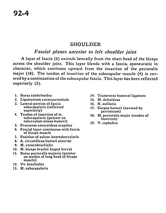
A layer of fascia (6)extends laterally from the short head of the biceps across the shoulder joint. This layer blends with a fascia, aponeurotic in character, which continues upward from the insertion of the pectoralis major (18). The tendon of insertion of the subscapular muscle (4) is covered by a combination of the subscapular fascia. This layer has been reflected superiorly (3).
- Subdeltoid bursa
- Coracoacromial ligament
- Lateral portion of subscapular fascia (reflected superiorly)
- Tendon of insertion of subscapularis muscle (pointer on lesser tubercle of humerus)
- Coracoid process of scapula
- Fascial layer continuous with fascia of biceps muscle
- Position of intertubercular sulcus
- Anterior circumflex artery of humerus
- Coracobrachialis muscle
- Biceps brachii muscle (short head)
- Pectoralis major bursa (pointer on tendon of long head of biceps muscle)
- Brachial veins
- Subscapularis muscle
- Transverse humeral ligament
- Deltoid muscle
- Axillary nerve
- Body of humerus (covered by periosteum)
- Pectoralis major muscle (tendon of insertion)
- Cephalic vein


