Bassett Collection of Stereoscopic Images of Human Anatomy
Pectoral region and axilla
Left subscapular muscle
Image #91-6
KEYWORDS: Axilla, Fascia ligaments and tendons, Pectoral region, Peripheral nervous system, Shoulder, Vasculature.
Creative Commons
Stanford holds the copyright to the David L. Bassett anatomical images and has assigned Creative Commons license Attribution-Share Alike 4.0 International to all of the images.

For additional information regarding use and permissions, please contact the Medical History Center.

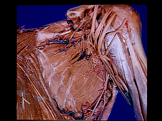
Pectoral region and axilla
Left subscapular muscle
The shoulder has been pulled away from the thoracic wall and the axillary artery and brachial plexus retracted laterally. The subscapular fascia (7) has been partially removed.
- Upper pointer: Clavicle (cut off) Lower pointer: Subclavius muscle (cut off)
- Transverse scapular artery (cut off)
- Suprascapular nerve (cut off)
- Omohyoid muscle (cut off)
- Medial angle of scapula
- Subscapularis muscle
- Subscapular fascia
- Serratus anterior muscle
- Long thoracic nerve
- Coracoid process of scapula
- Coracoclavicular ligament
- Lesser tubercle of humerus
- Subscapular nerves (upper subscapular nerves)
- Posterior cord of brachial plexus
- Axillary nerve
- Thoracodorsal nerve (middle subscapular nerve) (also see 21)
- Subscapular nerve (lower subscapular nerve)
- Coracobrachialis muscle
- Biceps brachii muscle
- Subscapular artery
- Thoracodorsal nerve (also see 16)
- Teres major muscle
- Latissimus dorsi muscle