Bassett Collection of Stereoscopic Images of Human Anatomy
Pectoral region and axilla
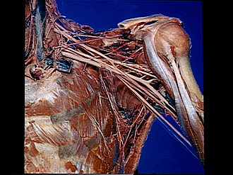
Left axilla (continued); brachial plexus and axillary artery
Image #91-4
KEYWORDS: Axilla, Peripheral nervous system.
Creative Commons
Stanford holds the copyright to the David L. Bassett anatomical images and has assigned Creative Commons license Attribution-Share Alike 4.0 International to all of the images.

For additional information regarding use and permissions, please contact the Medical History Center.


Pectoral region and axilla
Left axilla (continued); brachial plexus and axillary artery
The axillary vein and its branches have been cut away. Component parts of the brachial plexus have been separated slightly.
- Internal jugular vein
- Anterior scalene muscle
- Jugular trunk
- Subclavian artery
- Phrenic nerve
- Subclavian vein (cut off)
- Subscapularis muscle
- Rib II
- Long thoracic nerve
- Serratus anterior muscle
- Upper pointer: Cervical nerve V Lower pointer: Cervical nerve VI
- Cervical nerve VII
- Cervical nerve VIII
- Suprascapular nerve
- Transverse scapular artery
- Lateral cord of brachial plexus
- Branch axillary artery (to subscapularis muscle)
- Coracoid process of scapula
- Axillary nerve
- Musculocutaneous nerve
- Upper pointer: Coracobrachialis muscle Lower pointer: Anterior circumflex artery of humerus
- Posterior circumflex artery of humerus
- Axillary artery (note anomalous bifurcation a short distance distal to pointer. The lateral branch continues as the radial artery, the medial branch as the brachial artery)
- Radial nerve
- Thoracodorsal nerve
- Median nerve
- Biceps brachii muscle
- Ulnar nerve
- Middle antibrachial cutaneous nerve
- Thoracodorsal artery
- Teres major muscle
- Latissimus dorsi muscle